
Client: CHAPMAN TAYLOR s.r.o.
Field of activity: Chapman Taylor is an international architectural firm engaged in a wide range of projects, from urban district master plans to premium interiors. One of their latest projects in Prague is the interior of the recently opened W Hotel. Of their total 3,000 completed projects, a significant portion consists of office buildings and extensive fit-outs.
Contact/References
Ing. arch. Filip Pokorný, Ph.D., Director
fpokorny@chapmantaylor.com
Description of the problem or challenge:
We first encountered the renowned international firm Chapman Taylor in 2022 during their work on a major Prague renovation, where we introduced their team of architects to the benefits of full-spectrum lighting. Chapman Taylor was the chief architect of the project and was deeply involved in finding ways to improve the quality of the work environment and workplace lighting within a heritage-protected building in the center of Prague. As a result, the architects experienced firsthand the benefits our lighting provides—from accurate color rendering to a positive impact on work comfort.
In 2024, the architectural firm moved to new premises in the center of Prague, where natural lighting conditions were not ideal. The architects understand that proper lighting is crucial, and given their previous experience with our products in demanding projects for a major international developer, they contacted us for a customized solution.
Goals
- Provide architects with lighting that reduces eye strain and ensures maximum color accuracy.
- Enhance work comfort through optimized lighting.
- Improve overall health and well-being.
Solution description
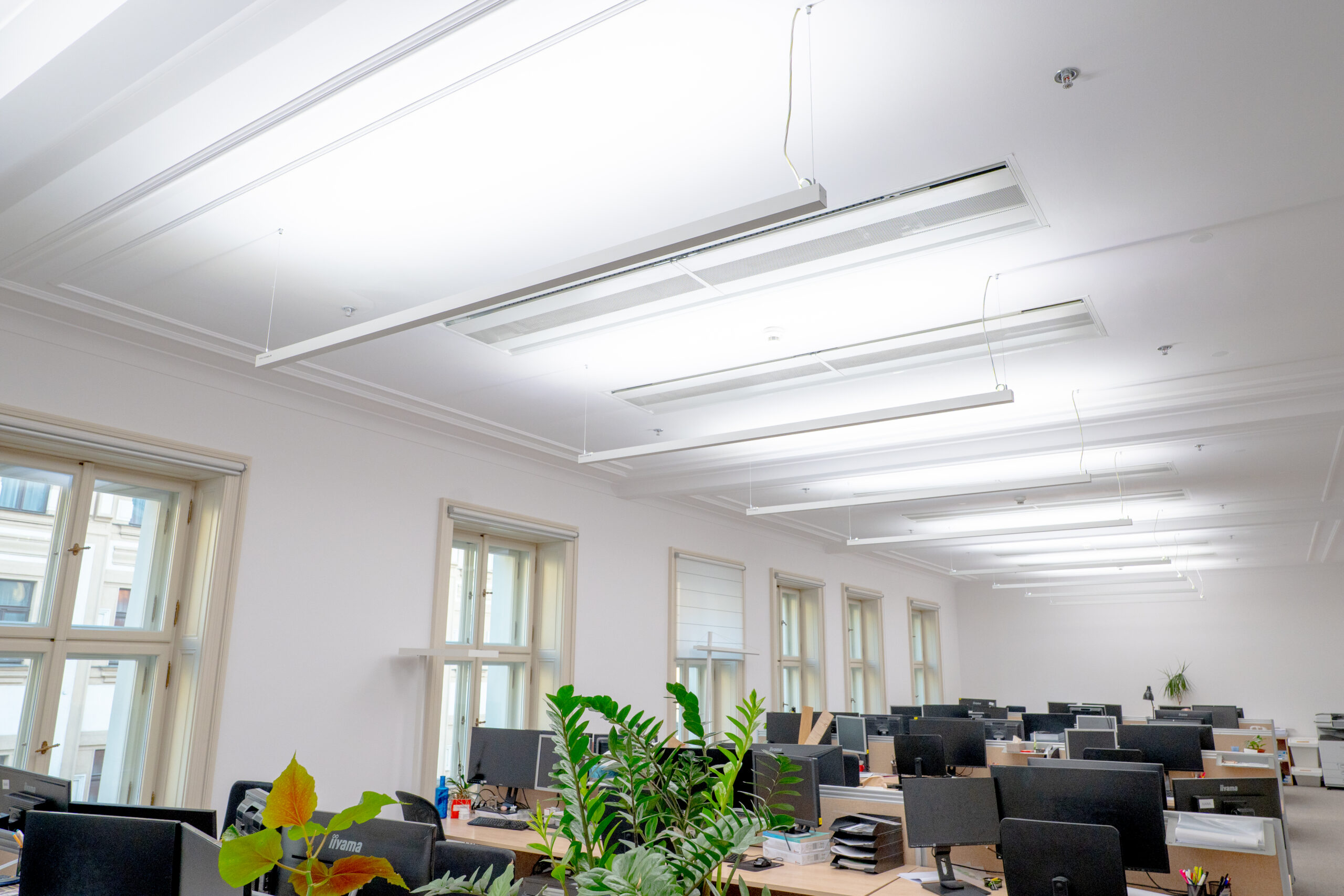
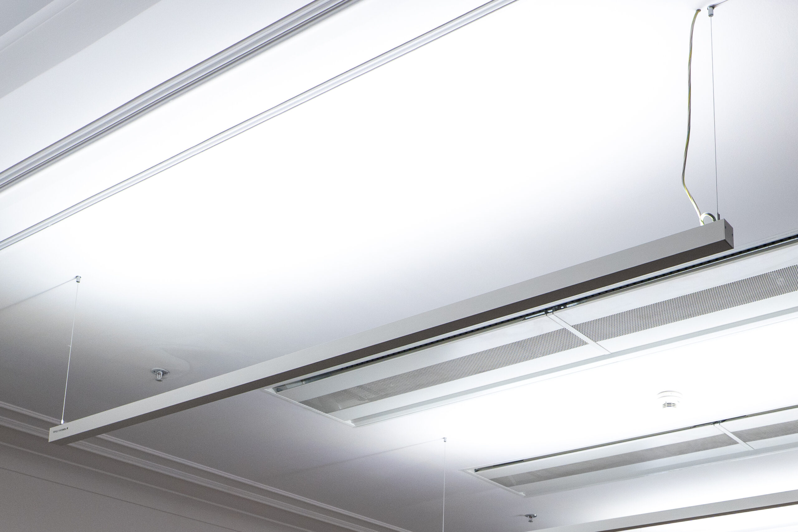
First, light-technical calculations had to be conducted, and a price proposal presented. The choice fell on the Sunline 45 linear indirect lighting system, which ensures not only excellent visual comfort but also significantly contributes to a pleasant atmosphere in the interior throughout the day.
Installation in the open space and meeting rooms of the office was completed within four weeks of the confirmed order.
Result:
The final outcome of the implementation is highly positive. The improvement in lighting quality was noticed not only by the client’s employees but also by their clients.
For example, a representative of a major Prague developer experienced something unexpected during a meeting at Chapman Taylor’s office: “When we were reviewing the newly prepared marketing visualizations with the architects, we were pleasantly surprised that the colors and details precisely matched the professionally calibrated monitors of the graphic designers. It was like discovering a whole new dimension of visualizations and being able to prepare them much more accurately for presentation!”
The architects themselves acknowledge the positive impact of the new lighting:
- Better focus – eye strain is significantly reduced due to consistent and natural light.
- Higher productivity – working with details is easier and faster.
- Natural color perception – accuracy is crucial for architectural designs.
And no more team disputes about whether to keep the office lights on during the day.
Client’s statement:
Managing director Ing. arch. Filip Pokorný, Ph.D., comments on the installation results:
“Consistent and natural lighting significantly reduces eye strain. We also observed excellent color accuracy, which is very important for architectural designs. Working with details has also become easier and faster, which enhances overall work productivity.”
Illustrative photo from the installation:
Translated using AI